Posts

CO-DOMINANCE ||Class 12 Biology

Image
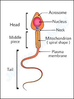
     In heterozygous condition both the alleles are expressed. Both alleles are expressed in dominant forms. The alleles which shows this property are called codominant alleles and the phenomenon is known as co-dominance.       In this condition, the offsprings shows resemblance to both the parents, eg- ABO blood groups in humans. ABO blood groups are controlled by the gene I. The plasma membrane of the red blood cells (RBCs) has sugar polymers that protrude from its surface and the kind of sugar is controlled by the gene. The gene (I) has three alleles: IA, IB and i. The alleles IA and IB produce a slightly different form of the sugar while allele i does not produce any sugar. Because humans are diploid organisms, each person possesses any two of the three I gene alleles. IA and IB are completely dominant over i, in other words when IA and i are present only IA expresses (because i does not produce any sugar) and when IB and i are present IB expresses. But...

Incomplete Dominance ||Class 12 Biology

Image
     Incomplete Dominance is defined by Carl Correns. It is a phenomenon in which the F1 hybrid shows intermediate characters of the parental genes. In incomplete dominance, the phenotypic ratio deviates from the normal Mendel's monohybrid ratio. Characters are not blending in F1 generation because parental types are recovered in F2 generation.       Example: Flowers colour in Mirabilis jalapa or 4 o' clock plant and Snapdragon or Antirrhinnus sp.         In a cross between red flower (RR) and white flower (rr) plant, the F1 generation obtained was pink (Rr). Again, when the same F1 progeny was self-pollinated, the F2 resulted in the ratio 1:2:1, i.e. not 3:1.

Pedigree Analysis ||Class 12 Biology

Image
     Pedigree analysis is an analysis of traits in several generations of a family. In this analysis, the inheritance of a particular trait is represented in the family tree over generations.         Importance of pedigree analysis are as follows:  1. It is used to know the possibility of expressive or recessive allele which may cause genetic disorders. 2. It shows the origin of trait and flow of that trait in a family. 3. It predicts the harmful effects of marriage between close relatives. 4. It is extensively used in medical research.       Pedigrees use a standardized set of symbols, squares represent males and circle represent females. It the sex of the person is unknown a diamond is used as symbol. Affected individuals is represented by a filled in (darker symbol). Heterozygotes, when identifiable, are indicated by a shade dot inside a symbol or a half filled symbol. Relationships in pedigree are shown as a series of lines...

Genetic Disorders ||Class 12 Biology

Image
    A number of disorders in human beings, are associated with the inheritance of changed and altered genes or chromosomes. These are called genetic disorders.  Since, it is not possible to perform control crosses in humans, study of family history about inheritance of a particular trait provides an alternative in the diagnosis of genetic disorders or abnormalities.       The genetic disorders can be classified as:  1. Mendelian disorders or Gene-related disorders 2. Chromosomal disorders 1. Mendelian disorders or Gene-related disorders: These are the disorders that are mainly determined by alterations or mutation in the single gene. These disorders are transmitted in the next generation according to the principle of inheritance and can be studied by the pedigree analysis. The diseases may occur as dominant or recessive. Example: Haemophilia, colour blindness, sickle-cell anaemia, phenylketonuria etc. 2. Chromosomal disorder: These are caused by the ab...

Colour Blindness: Causes & Symptoms ||Class 12 Biology

Image
      Colour blindness is a recessive sex-linked trait. The recessive allele are carried by X-chromosome. Colour blindness is also known as colour vision deficiency, is the decreased ability to see differences in colour. Red-green colour blindness is the most common form, followed by blue-yellow colour blindness and total colour blindness. In Red-green colour blindness, the affected eye fail to distinguish between red and green colours.      The ishihara colour test, which consists of a series of pictures of coloured spots, is the test most often used to diagnose red-green colour deficiencies. Ishihara card       In female, it appears only when both the sex chromosome carry the gene (XcXc). The females function as carries in the presence of a single recessive gene (XXc) while in males the defects appears in the presence of a single recessive gene (XcY) because Y-chromosome does not carry any gene for colour vision.      There i...

Placenta: Structure & function ||Class 12 Biology

Image
     The placenta is an organ attached to the lining of womb during pregnancy. Structure:       After implantation, finger-link projections appears on the trophoblast called chorionic villi which surrounded by the uterine tissues and maternal blood. The chorionic villi and the uterine tissue become interdigitated with each other and jointly form a structural and and functional unit between foetus and maternal body called placenta. Placenta is forms from the chorian and allantois. In human, the placenta averages 22cm (9 inch) in length and 2-2.5cm (0.8-1inch) in thickness. it has dark reddish-blue or crimson colour. It connects to the foetus by an umbilical cord. Function:      1. It facilitates the supply of oxygen and nutrients to the embryo.     2. Responsible for the removal of carbon dioxide and waste materials produced by the foetus.    3. The placenta is connected to the embryo through the umbilical cord which h...

Antibody: Structure ||Class 12 Biology

Image
      Antibodies are immunoglobulin (Ig)  molecules. An Antibody seems like a Y-shaped structure. Antibodies are of mainly 5 types such as, IgA, IgM, IgE, IgG and IgD.       Antibodies are glycoproteins. They are typically made of basic structural units that consists of four polypeptide chains: two identical large heavy (H) chains and two identical small light (L) chains connected by disulphide bonds. Hence, an Antibody is represented as H 2 L 2 . The size of an antibody molecule is about 10nm. Each of the four chains has two regions: the constant region and the variable region.         The antibody recognizes a unique molecule of the pathogen, called an antigen via fragment antigen-binding (Fab) site of variable region. An antigen binding site is also called paratope, is a part of an antibody which recognizes and binds to an antigen. The part of antigen to which the paratope binds is called an epitope. The ability of an antibody to...

Sex Determination in Humans (XX-XY type) ||Class 12 Biology

Image
     In humans, 23 pairs of chromosomes are present out of which 22 pairs are exactly same in both males and females, these are autosomes. The 23rd pair of chromosome is called sex chromosome.       A pair of X-chromosome (XX) is present in females, whereas one X and one Y chromosome (XY) is present in males. In males, during Spermatogenesis, two types of gametes are produced. 50% of the total sperm produced carry X chromosome and the rest 50% carry Y chromosomes besides autosomes. The female produces only one type of ovum with an X-chromosome.      In case, the ovum fertilises with a sperm carrying X-chromosome, the zygote develops into a female (XX) and if ovum is fertilised with sperm carrying Y-chromosome, zygote formed will be a male (XY). Thus, the genetic makeup of sperm which fertilises an ovum determines the sex of a child. So, women should not be blamed for the sex of a child. There are total 50% chances of having a male child ...

Sperm: Structure ||Class 12 Biology

Image
     A mature spermatozoa or a sperm cell is a microscopic, tadpole-shaped structure of about 2-5 micrometer in diameter and 50 micrometer long. A plasma membrane encloses the whole body of sperm.      Sperm consists of head, neck, middle piece and tail.       1. Head: It is flat, oval and composed of a large haploid nucleus and a small cap-like structure called acrosome. It lies at the tip of the nucleus and is formed from the Golgi complex. It contains hydrolytic enzymes and is used to contact and penetrate the egg (ova) during fertilization.       2. Neck: Neck is the smallest part, it is short structure that contains two centrioles- one parallel to the base of the nucleus and called proximal centriole and other distal centriole perpendicular to the previous one. The proximal centriole enters into the egg during fertilization and starts the first cleavage division of the egg which has no centriole. The distal centriole give...

Turner's Syndrome: Causes & Symptoms ||Class 12 Biology

Image
      Turner's syndrome is a sex chromosomal abnormality. It is caused due to the absence of one of the X chromosome i.e. 45 with XO in which a female is partly or completely missing an X chromosome.        Turner's syndrome was named after Henry Turner, an endocrinologist who described it in 1938. Turner's syndrome occurs in between 1 in 2000.       Affected females are sterile as ovaries are rudimentary, i.e. undeveloped. Lack of secondary sexual characters and poor breasts development. Short stature, small uterus, puffy fingers, webbed neck, swollen hands and feet are seen at birth.            Turner's syndrome is not usually inherited in families. The exact cause of Turner's syndrome is not known. It appears to occur as a result of a random error during the formation of either eggs or sperms.       No cure for Turner's syndrome is known. Treatment may help with symptoms. Human Growth ...